
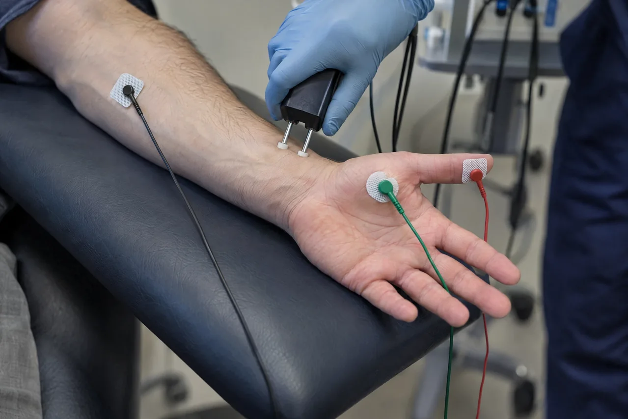
The study & its electrophysiologic principle
Surface electrodes stimulate a selected motor nerve and record a compound muscle action potential (CMAP) from a target muscle. Trains are repeated under controlled conditions, sometimes before and after brief activation. A reproducible decrement or post-activation increment may indicate transmission dysfunction; interpretation depends on muscle selection and technical quality.
RNS tests the safety margin of synaptic transmission. Low-rate stimulation may expose postsynaptic failure, whereas activation or faster stimulation can transiently increase presynaptic acetylcholine release in some disorders. Temperature, electrode stability, activation, medications, and laboratory reference methods affect results.
Common clinical applications
RNS is commonly used when evaluation raises concern for neuromuscular transmission disorders, including suspected myasthenia gravis or presynaptic syndromes such as Lambert-Eaton myasthenic syndrome. Patterns may suggest postsynaptic or presynaptic physiology, but sensitivity varies with disease distribution, severity, medicines, and muscle tested.
It can complement NCS, needle EMG, antibody or other laboratory testing, and neurologic examination in fluctuating, fatigable, ocular, bulbar, limb, or proximal weakness. A normal study does not exclude disease, and an abnormal response is not synonymous with a named diagnosis.
What you may expect
Small surface electrodes are placed over a nerve and muscle. Brief pulses may feel like tapping, tingling, or a quick twitch; discomfort varies, and a painless test cannot be promised. Needles are not used unless a separately indicated needle EMG is performed.
Several short recording series may be collected, sometimes after brief muscle activation. Visit length and result timing vary by protocol and service, so immediate results or a particular outcome cannot be guaranteed.
Wear comfortable clothing and keep electrode sites free of lotions or oils. Tell the team about medicines, supplements, implanted devices, bleeding concerns, and relevant conditions. Do not stop or change prescribed medicines unless your managing clinician instructs you.
Explore the technical detail.
Open the sections below for anatomy, physiologic parameters, methodology and clinical limitations.
Anatomical & physiologic context
The pathway runs from peripheral motor nerve stimulation through the neuromuscular junction to muscle fibers whose combined response is recorded at the skin. Muscles are selected according to the clinical question; facial and limb muscles may differ in recording characteristics and sensitivity.
RNS is distinct from conventional sensory and motor conduction measurements and from needle EMG. It emphasizes the behavior of successive evoked motor responses under controlled stimulation and activation conditions.
Important physiologic parameters
- CMAP amplitude, area, configuration, and latency across successive stimuli.
- Reproducible decrement at low rates or increment after activation or faster rates, judged across traces rather than one response.
- Stimulation frequency, train length, intensity, timing of activation, and post-activation recovery.
- Skin temperature, muscle relaxation or immobilization, electrode placement, artifact control, and reproducibility.
- Comparison with laboratory-specific reference procedures and the clinically relevant muscle; no universal cutoff should be applied outside its validated context.
General methodology
An appropriately trained professional prepares the skin, places surface electrodes, stabilizes the muscle when appropriate, and verifies a supramaximal response. Protocol-defined trains are recorded at selected rates; activation and recovery measurements may be added. Temperature and artifact are controlled because they alter responses.
RNS is data acquisition. Licensed medical interpretation, diagnostic correlation, and treatment decisions belong to the responsible qualified clinician under applicable law and arrangements. Separately requested needle EMG requires an appropriately qualified licensed professional and is not replaced by RNS.
Clinical relevance & limitations
Findings are interpreted with symptom history, neurologic examination, muscle selection, medication and activation conditions, temperature, technical quality, and other studies. Data complement, not replace, clinical assessment and do not by themselves provide a definitive diagnosis or determine treatment.
The recorded responses support clinical evaluation within the appropriate study framework. The responsible licensed clinician determines their medical significance and whether additional investigation is needed.
- A negative or technically limited RNS cannot rule out neuromuscular-junction disease; CMAP change is nonspecific and requires validated laboratory methods and clinical correlation.
- RNS does not assess every cause of weakness. NCS and needle EMG answer different questions, while blink reflex, F-wave, and H-reflex studies are separate physiologic tests.
Information, within appropriate scope.
Recorded physiologic data support broader clinical evaluation. Licensed medical interpretation, diagnosis and treatment are separate clinical responsibilities. Specific service availability, professional qualifications and arrangements must be verified.
Educational references
Selected sources support the educational discussion. They do not imply affiliation, endorsement or an individual clinical recommendation.
- Practice parameter for repetitive nerve stimulation and single fiber EMG evaluation of adults with suspected myasthenia gravis or Lambert-Eaton myasthenic syndrome (opens in a new tab)
- Disorders of Neuromuscular Transmission (opens in a new tab)
- Myasthenia gravis: Diagnosis and treatment (opens in a new tab)
- Myasthenia Gravis (opens in a new tab)