
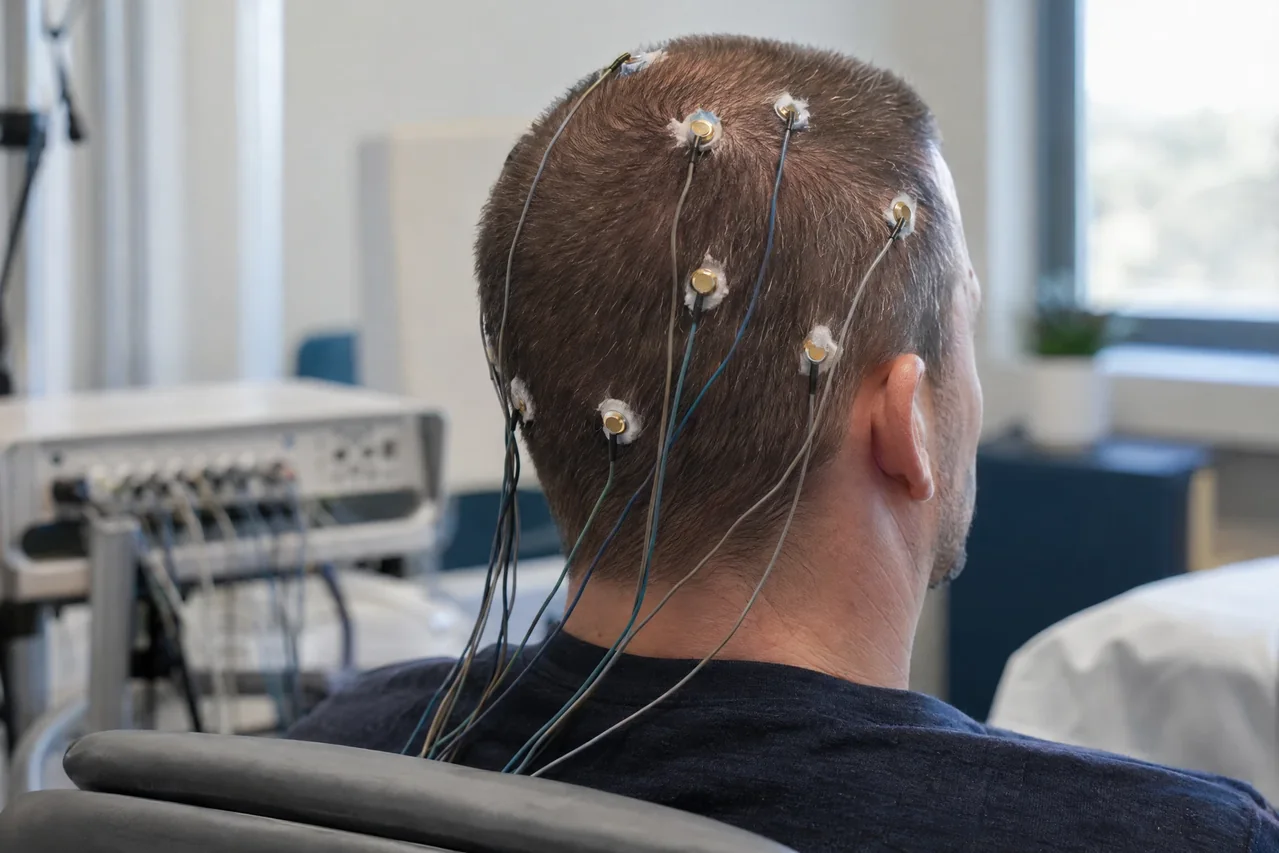
The study & its electrophysiologic principle
Scalp electrodes detect tiny potential differences, which are amplified, digitized, displayed, and stored as multichannel waveforms. The signal chiefly reflects summed postsynaptic activity from synchronized cortical neuronal populations, filtered by tissue, skull, and scalp. EEG has excellent temporal resolution but limited spatial specificity compared with imaging.
Interpretation considers background, state, reactivity, transients, sleep features, artifacts, and events captured with video or other channels. Eye, muscle, cardiac, electrode, and electrical interference can mimic cerebral activity. EEG complements history, examination, imaging, laboratory data, and other studies; it cannot establish a definitive diagnosis alone.
Common clinical applications
EEG commonly evaluates suspected seizures and epilepsy, paroxysmal events, encephalopathy or altered consciousness, and selected sleep-related or critical-care questions. Routine, sleep, ambulatory, and prolonged video-EEG designs differ in their opportunity to capture events.
EEG may contribute to assessment of cerebral dysfunction in selected toxic-metabolic, infectious, structural or other clinical contexts. Indication, acquisition protocol and interpretation depend on the clinical question and the responsible clinician’s judgment.
What you may expect
EEG uses recording electrodes rather than electrical stimulation, so electrodes do not send current into the brain. Most people feel little or no discomfort, although preparation, paste, and removal can irritate; complete absence of discomfort cannot be guaranteed.
You may be asked to rest, open or close the eyes, breathe in a specified way, view flashing light, or sleep. Setup and recording time varies; immediate results and a particular outcome should not be assumed.
General preparation commonly includes clean, dry hair without styling products and following the ordering clinician’s instructions about sleep or food. Do not stop, change, or withhold medicines unless directed; disclose medicines and conditions. If sedation is considered, the responsible team should explain safety and transportation.
Explore the technical detail.
Open the sections below for anatomy, physiologic parameters, methodology and clinical limitations.
Anatomical & physiologic context
A scalp array samples frontal, central, temporal, parietal, and occipital regions using an organized 10–20-type placement system or high-density montage. Channels compare potentials in a defined montage, so waveform distribution and polarity partly depend on reference and display configuration.
EEG is most sensitive to sufficiently synchronous cortical activity that can be detected at the scalp; deep or spatially restricted sources may be poorly represented. It records functional electrical activity rather than producing a structural image of the brain.
Important physiologic parameters
- Background frequency, amplitude, continuity, symmetry, and organization during wakefulness and sleep.
- Reactivity to eye opening, eye closure, arousal, and appropriate activation procedures.
- Distribution, morphology, persistence, and state dependence of rhythmic or epileptiform-appearing transients.
- Focal versus generalized slowing or other abnormalities, with montage, field, and artifact assessment.
- Timing of electrographic changes relative to symptoms, behavior, video, ECG, EOG, or EMG channels when acquired.
- Technical quality, electrode contact, sampling, filtering, montage, annotations, and recording state.
General methodology
A trained technologist prepares the scalp, applies paste or gel, attaches electrodes or a cap, checks signal quality, and records specified channels. The patient may relax, open or close the eyes, breathe in a specified way, view flashing light, or sleep, depending on the protocol.
Acquisition is technical data collection; licensed medical interpretation is separate. A qualified interpreter reviews the recording and clinical context and communicates findings through the responsible healthcare pathway.
Clinical relevance & limitations
An EEG finding gains meaning from the indication, symptoms, examination, medication and sleep state, technical quality, and other investigations. A captured electrographic event can be informative, but an event without a characteristic EEG correlate and an abnormal waveform are not automatically a clinical seizure or specific disease.
A report is a medical interpretation, not a standalone verdict. A normal or nondiagnostic study may reflect limited sampling or absence of an abnormality during recording; slowing and artifacts require context. The treating clinician determines how results fit the individual’s care.
- EEG acquisition does not itself diagnose, treat, prescribe, or replace licensed interpretation; findings are complementary evidence and do not independently establish a diagnosis.
- A normal or nondiagnostic recording does not exclude every clinically relevant disorder; sampling period, patient state, technical quality and source location influence what is recorded.
Information, within appropriate scope.
Recorded physiologic data support broader clinical evaluation. Licensed medical interpretation, diagnosis and treatment are separate clinical responsibilities. Specific service availability, professional qualifications and arrangements must be verified.
Educational references
Selected sources support the educational discussion. They do not imply affiliation, endorsement or an individual clinical recommendation.