
The study & its electrophysiologic principle
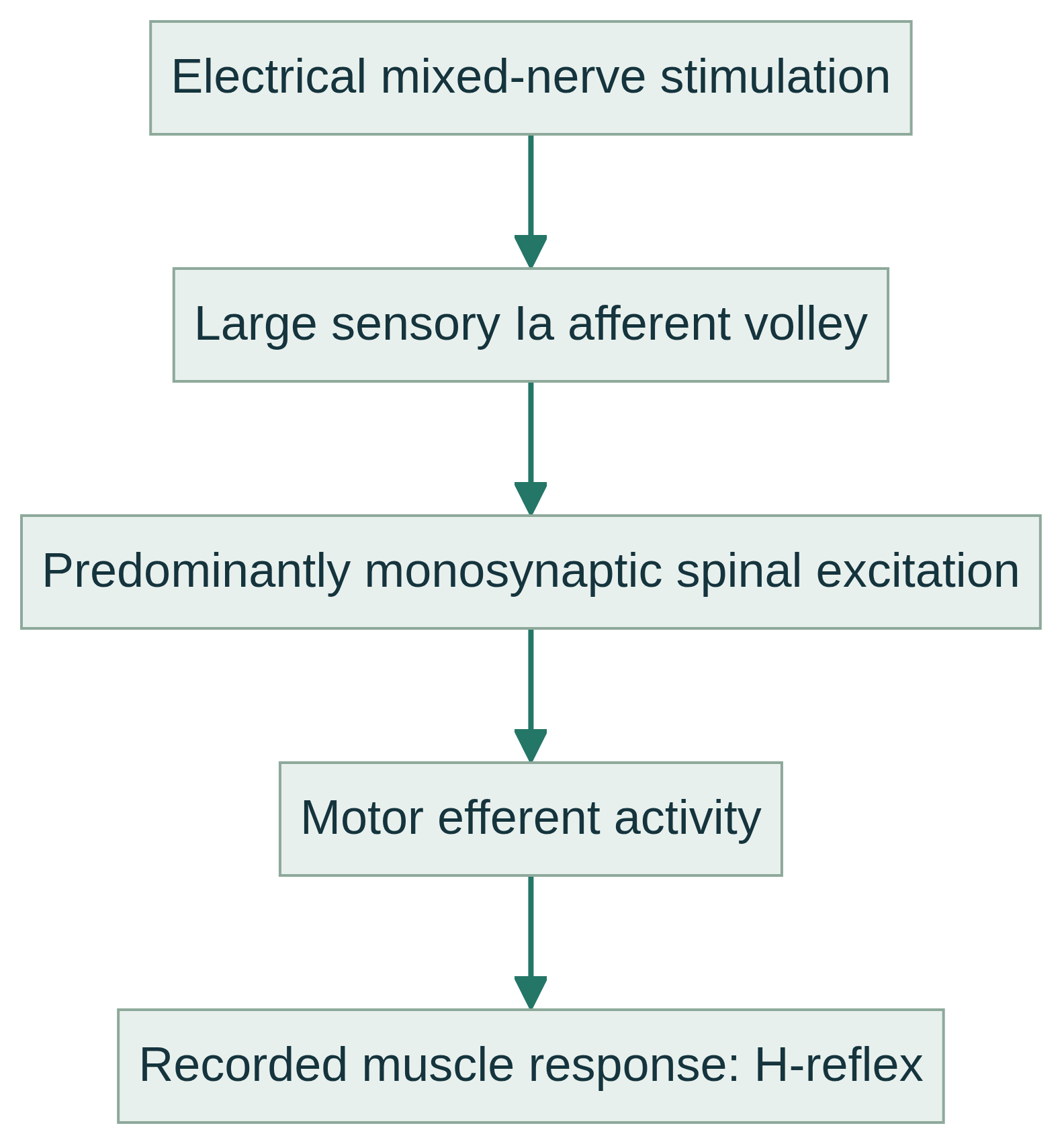
A surface stimulus to a mixed nerve preferentially recruits large, low-threshold sensory Ia afferents. Their volley travels to the spinal cord, engages predominantly monosynaptic excitation of motor neurons, and returns through motor axons to produce a muscle response. Direct afferent activation bypasses the muscle-spindle receptor and mechanical stretch.
The response is predominantly, not exclusively, monosynaptic: oligosynaptic inputs and presynaptic and motor-neuron properties influence its size. It is not a standalone measure of motor-neuron-pool excitability or a diagnosis. An F-wave is a different late motor response after supramaximal stimulation and antidromic motor-axon activation; a blink reflex examines trigeminal-afferent, facial-efferent, and brainstem circuitry.
Common clinical applications
When indicated, H-reflexes can provide complementary information in suspected S1 or other radicular dysfunction, sciatic or sacral-plexus involvement, proximal mononeuropathy, or large-myelinated-fiber disorders. An abnormal or absent response is not specific to one lesion.
Findings may contribute to assessment of selected polyneuropathies, multifocal conduction abnormalities, or proximal nerve disease. A qualified medical examiner selects muscles, nerves, sides, and companion studies and integrates results with history, examination, NCS, and indicated needle EMG.
What you may expect
The team will explain the procedure, place surface electrodes and ask you to relax or maintain an appropriate position. Duration depends on the recording protocol and comparisons required; immediate results should not be assumed.
You may feel short electrical pulses, tingling, pressure, or brief involuntary movement. Most people tolerate surface stimulation, yet pain or discomfort cannot be guaranteed absent. Mention relevant symptoms, implanted electrical devices, or skin concerns before testing.
General preparation is simple: wear clothing permitting limb access, keep electrode sites free of lotion, and follow site instructions. Do not stop, change, or omit medicines unless the licensed clinician managing them instructs you to do so.
Explore the technical detail.
Open the sections below for anatomy, physiologic parameters, methodology and clinical limitations.
Anatomical & physiologic context
The circuit spans a stimulated mixed nerve, large myelinated sensory afferents, spinal segments and connections, motor axons, and the recorded muscle. In adults, soleus responses commonly sample tibial-nerve and S1-region pathways; flexor carpi radialis responses can sample upper-limb C6–C7-region pathways. Muscle and side selection follows the clinical question.
Important physiologic parameters
- Response latency: stimulus-to-wave interval reflecting conduction and synaptic timing.
- Response amplitude: usually peak-to-peak; it reflects recruited synchronous activity but is sensitive to technical and physiologic conditions.
- Waveform reproducibility and morphology: consistency across trials supports technical quality.
- H-to-M relationship: comparison with the directly evoked M-wave helps document stimulus intensity and contextualize recruitment, not establish a universal cutoff.
- Side-to-side comparison: asymmetry may be informative but requires attention to anatomy, temperature, age, limb length, and method.
- Recording conditions: nerve and muscle tested, stimulus site and intensity, electrode placement, posture, temperature, and muscle activation materially affect measurements.
General methodology
After skin preparation, surface recording, reference, and ground electrodes are placed as appropriate. Brief electrical stimuli are delivered over the nerve, with repeated trials and controlled conditions used to characterize the response. Sensation is usually brief tingling or tapping with possible muscle contraction, but discomfort cannot be guaranteed absent.
H-reflex acquisition evaluates a different physiologic response from conventional sensory and motor conduction measurements, although they may be acquired within one broader examination. Needle EMG is a separate procedure requiring appropriately qualified licensed professional performance. Acquisition does not itself constitute medical interpretation, diagnosis or treatment.
Clinical relevance & limitations
An H-reflex result is one physiologic observation, not a definitive diagnosis. Latency, amplitude, absence, or asymmetry can reflect lesion location, technique, temperature, body habitus, age, medications, activation state, or biologic variability. A licensed professional interprets the complete study with history, examination, other electrodiagnostic data, and relevant imaging or laboratory information.
Apex NeuroMetrics distinguishes measurement from medical decision-making: acquired data support clinical evaluation, while licensed interpretation, diagnosis, and treatment decisions do not arise from a waveform alone. Questions about findings belong with the licensed professional responsible for evaluation.
- H-reflexes are technically and physiologically sensitive; reference ranges vary with muscle, method, population, temperature, and laboratory technique. No single cutoff or isolated result diagnoses disease.
- H-reflex studies complement rather than replace history, examination, NCS, and indicated needle EMG. An H-reflex alone cannot establish radiculopathy, neuropathy, treatment need, or prognosis.
Information, within appropriate scope.
Recorded physiologic data support broader clinical evaluation. Licensed medical interpretation, diagnosis and treatment are separate clinical responsibilities. Specific service availability, professional qualifications and arrangements must be verified.
Educational references
Selected sources support the educational discussion. They do not imply affiliation, endorsement or an individual clinical recommendation.
- The H-reflex as a tool in neurophysiology: its limitations and uses in understanding nervous system function (PubMed, University of Alberta review) (opens in a new tab)
- Basic Electrodiagnostics for the Referring Physician (American Academy of Physical Medicine and Rehabilitation) (opens in a new tab)
- The H-reflex as a tool in neurophysiology: its limitations and uses in understanding nervous system function (University of North Carolina Biomedical Engineering hosted review) (opens in a new tab)