
The study & its electrophysiologic principle
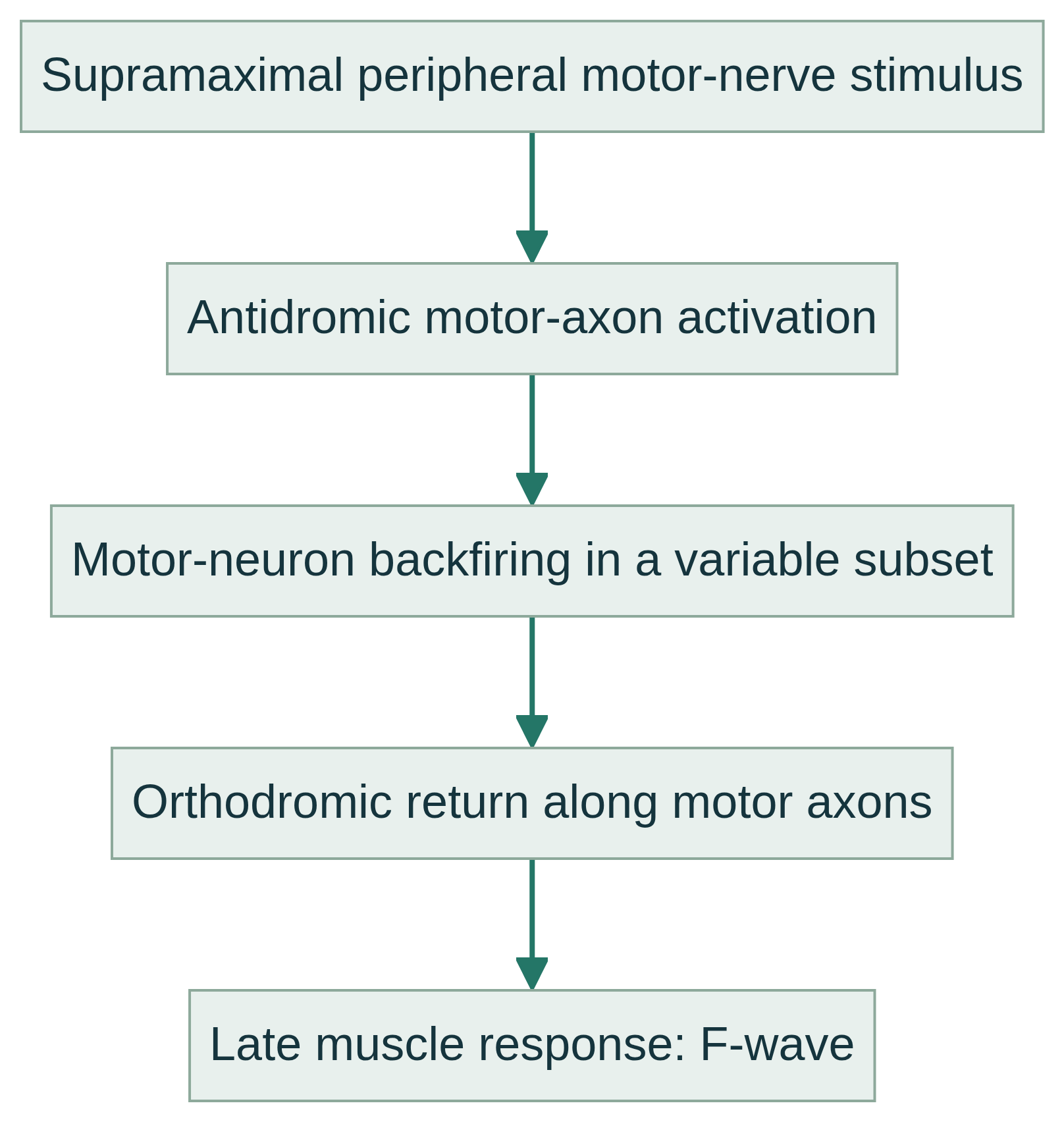
Surface electrodes deliver a brief electrical stimulus to a peripheral motor nerve while recording from a target muscle. Besides the immediate M wave, some antidromically activated motor axons send an impulse toward the spinal motor neuron pool; a small, variable group then backfires orthodromically to produce the late F wave. F-waves are late motor responses, not reflexes, and may be absent on individual trials.
Because the impulse traverses much of the nerve twice, the response can provide information about distal and proximal segments, including regions near roots and plexus less accessible to conventional stimulation. Repeated stimuli are used because latency, amplitude, configuration, and occurrence vary. Interpretation requires suitable technique, reference data, and correlation with the electrodiagnostic examination.
Common clinical applications
F-waves can complement routine motor and sensory nerve conduction studies when evaluating suspected generalized peripheral neuropathy, proximal mononeuropathy, plexopathy, or proximal or multifocal conduction abnormalities. They may contribute evidence in suspected inflammatory demyelinating neuropathy or Guillain–Barré syndrome, but no single F-wave feature establishes these conditions.
F-waves have limited specificity for focal root lesions because each response samples a subset of motor axons along a long pathway. Suspected radicular dysfunction requires integration with the examination and other appropriate electrodiagnostic components, including qualified needle EMG when indicated.
What you may expect
You will usually sit or lie comfortably while adhesive surface electrodes are placed on the skin and a nerve is stimulated. Pulses can feel startling, tapping, or briefly uncomfortable, and muscle twitching may occur; sensation varies and complete absence of discomfort cannot be guaranteed.
Repeated brief stimuli sample variable late responses. Time required depends on the nerves and clinical questions, so a fixed duration or immediate result should not be assumed. F-wave data are considered with other NCS findings and the clinical record.
General preparation is usually simple: wear clothing permitting limb access, keep electrode sites free of lotion, and report relevant conditions, implanted electrical devices, medications, and symptoms. Do not stop or alter prescribed medication unless a qualified clinician who knows your care specifically instructs you to do so.
Explore the technical detail.
Open the sections below for anatomy, physiologic parameters, methodology and clinical limitations.
Anatomical & physiologic context
The pathway follows large motor axons from the stimulated peripheral nerve toward the spinal motor neuron pool and back to the target muscle. The nerve and muscle depend on the clinical question. F-wave physiology differs from the H-reflex, a predominantly monosynaptic Ia-afferent, spinal-synapse, alpha-motor-efferent pathway; it also differs from the blink reflex, involving trigeminal input, facial motor output, and brainstem connections.
Important physiologic parameters
- Minimum onset latency: earliest observed F-wave onset after stimulation.
- Maximum onset latency: latest observed onset in the sampled responses.
- Latency dispersion or chronodispersion: range between minimum and maximum onset latency.
- Persistence: proportion of stimuli producing an identifiable F-wave; it is inherently variable.
- Amplitude and waveform configuration: small, variable features assessed alongside latency and persistence, not as standalone proof of disease.
General methodology
After skin preparation, surface recording electrodes are placed over the muscle motor point, with a reference electrode over a distal tendon or inactive bony site and a ground between recording and stimulation sites. The nerve is stimulated percutaneously with brief pulses increased to supramaximal intensity, while multiple responses are acquired with an extended recording window.
The operator documents the nerve, stimulation and recording sites, stimulus conditions, limb temperature, and responses. F-wave acquisition is part of NCS and is not needle EMG. Needle EMG, when indicated, requires an appropriately qualified licensed professional; data acquisition alone is not medical interpretation, diagnosis, or treatment.
Clinical relevance & limitations
Results can be influenced by limb length, age, temperature, electrode placement, motor axon excitability, and recording factors. Interpretation uses validated, technique-appropriate reference information and clinical context rather than universal cutoff values.
Apex NeuroMetrics educational information does not replace licensed medical interpretation. Test data complement history, neurological examination, routine NCS, needle EMG when indicated, and other investigations; they are never a definitive diagnosis by themselves. Diagnosis and treatment decisions remain with the responsible clinician.
- A normal, prolonged, low-persistence, or absent F-wave is not independently diagnostic.
- F-wave acquisition is distinct from needle EMG, H-reflex, and blink-reflex testing; the appropriate qualified licensed professional must determine, perform, and interpret any broader evaluation.
Information, within appropriate scope.
Recorded physiologic data support broader clinical evaluation. Licensed medical interpretation, diagnosis and treatment are separate clinical responsibilities. Specific service availability, professional qualifications and arrangements must be verified.
Educational references
Selected sources support the educational discussion. They do not imply affiliation, endorsement or an individual clinical recommendation.
- American Association of Neuromuscular & Electrodiagnostic Medicine. Recommended Policy for Electrodiagnostic Medicine (2023) (opens in a new tab)
- Association of Neurophysiological Technologists of Australia. ANTA Inc. NCS Guideline (2016) (opens in a new tab)
- Jerath N, Kimura J. F wave, A wave, H reflex, and blink reflex. Handbook of Clinical Neurology (2019); PubMed record (opens in a new tab)