
The study & its electrophysiologic principle
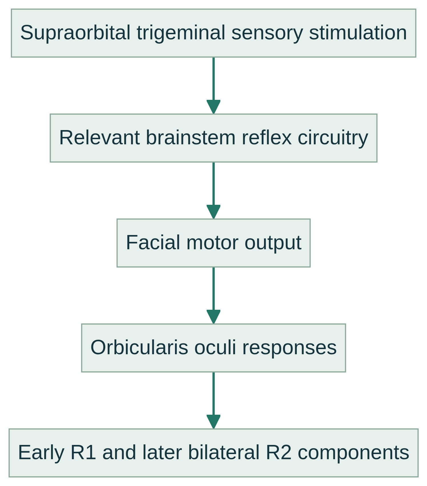
The study applies brief controlled electrical stimuli to a trigeminal sensory territory while surface electrodes record the orbicularis oculi. Responses are commonly described as R1, an earlier usually ipsilateral component, and R2, a later often bilateral component. Presence, latency, reproducibility, and side-to-side relationships inform assessment of afferent, brainstem, and facial efferent pathways.
Blink responses are reflexes, not voluntary blinks. Trigeminal input reaches pontine and medullary interneuronal networks, which activate facial motor nuclei and orbicularis oculi. The study is distinct from limb nerve conduction studies and needle EMG. F-waves are late motor responses after supramaximal stimulation, not reflexes; an H-reflex predominantly tests monosynaptic afferent, spinal, and efferent physiology.
Common clinical applications
Clinicians may request testing for suspected trigeminal or facial nerve dysfunction, selected brainstem lesions, facial neuropathy, trigeminal disorders, or other clinically relevant trigeminal–facial circuit questions. It is not a stand-alone disease test.
The study can complement examination, history, imaging, routine nerve conduction studies, and other electrodiagnostic findings, including selected cranial neuropathy or brainstem evaluations. Protocol and value depend on the suspected lesion, timing, and context.
What you may expect
You generally sit or recline while small surface electrodes are placed near the eyes and brief electrical stimulation is delivered to the face. It may feel startling, tingling, or uncomfortable; no test can guarantee painless sensations.
Brief eyelid or facial contractions may occur. Relaxing, keeping still, and following instructions support usable recordings. The procedure does not promise immediate results, a particular duration, or a particular outcome.
For general preparation, arrive with clean skin and report relevant conditions, eye or facial concerns, and medicines or devices that may affect testing. Do not stop or change medicines unless your responsible clinician gives individualized instructions.
Explore the technical detail.
Open the sections below for anatomy, physiologic parameters, methodology and clinical limitations.
Anatomical & physiologic context
The circuit includes ophthalmic trigeminal sensory fibers, trigeminal brainstem nuclei and interneurons, facial motor nuclei, and facial nerves to orbicularis oculi. Bilateral brainstem connections mean stimulation on one side can produce responses in both muscles.
Recordings sample a distributed trigeminal–facial brainstem network, not one nerve segment. Eye condition, facial activation, alertness, stimulus placement, and technique can affect responses.
Important physiologic parameters
- Stimulus site and side: trigeminal territory, laterality, electrode placement, and stimulus delivery are documented.
- R1 latency and reproducibility: timing and consistency of the early, predominantly ipsilateral response.
- R2 latency and reproducibility: timing and consistency of the later ipsilateral and contralateral responses.
- Presence and morphology: identifiable components, waveform stability, duration, and recording quality.
- Side-to-side comparison: relative latency, symmetry, and absent or delayed components, considered with technical conditions.
- Interstimulus timing when relevant: stimulus history can alter reflex excitability and must be interpreted within protocol.
General methodology
A qualified testing professional prepares the skin, places surface electrodes over orbicularis oculi, delivers brief electrical stimuli to a trigeminal site, and records repeated responses. Stimulus and recording conditions, artifact, movement, muscle tension, and relaxation are monitored.
Blink reflex acquisition produces response waveforms and measurements, not a diagnosis. Qualified clinical interpretation integrates these findings with the examination and other relevant information. Needle EMG, if separately indicated, is a distinct procedure requiring appropriately qualified licensed professional performance.
Clinical relevance & limitations
Interpretation considers response patterns, age, technical conditions, side-to-side comparison, symptoms, examination, disease timing, and other studies. Delayed, asymmetric, or absent responses have multiple possible peripheral, brainstem, technical, or physiologic explanations.
Blink data complement—not replace—history, neurologic examination, imaging, nerve conduction studies, and other electrodiagnostic tests. Data acquisition does not confer authority to diagnose or treat; a licensed medical professional interprets findings within the clinical record.
- No single blink response, latency, or waveform establishes a definitive diagnosis; normative limits vary with protocol, equipment, recording conditions, and patient factors.
- The study does not independently determine treatment, prognosis, or symptom cause; licensed medical interpretation and clinical correlation are required.
Information, within appropriate scope.
Recorded physiologic data support broader clinical evaluation. Licensed medical interpretation, diagnosis and treatment are separate clinical responsibilities. Specific service availability, professional qualifications and arrangements must be verified.
Educational references
Selected sources support the educational discussion. They do not imply affiliation, endorsement or an individual clinical recommendation.