
The study & its electrophysiologic principle
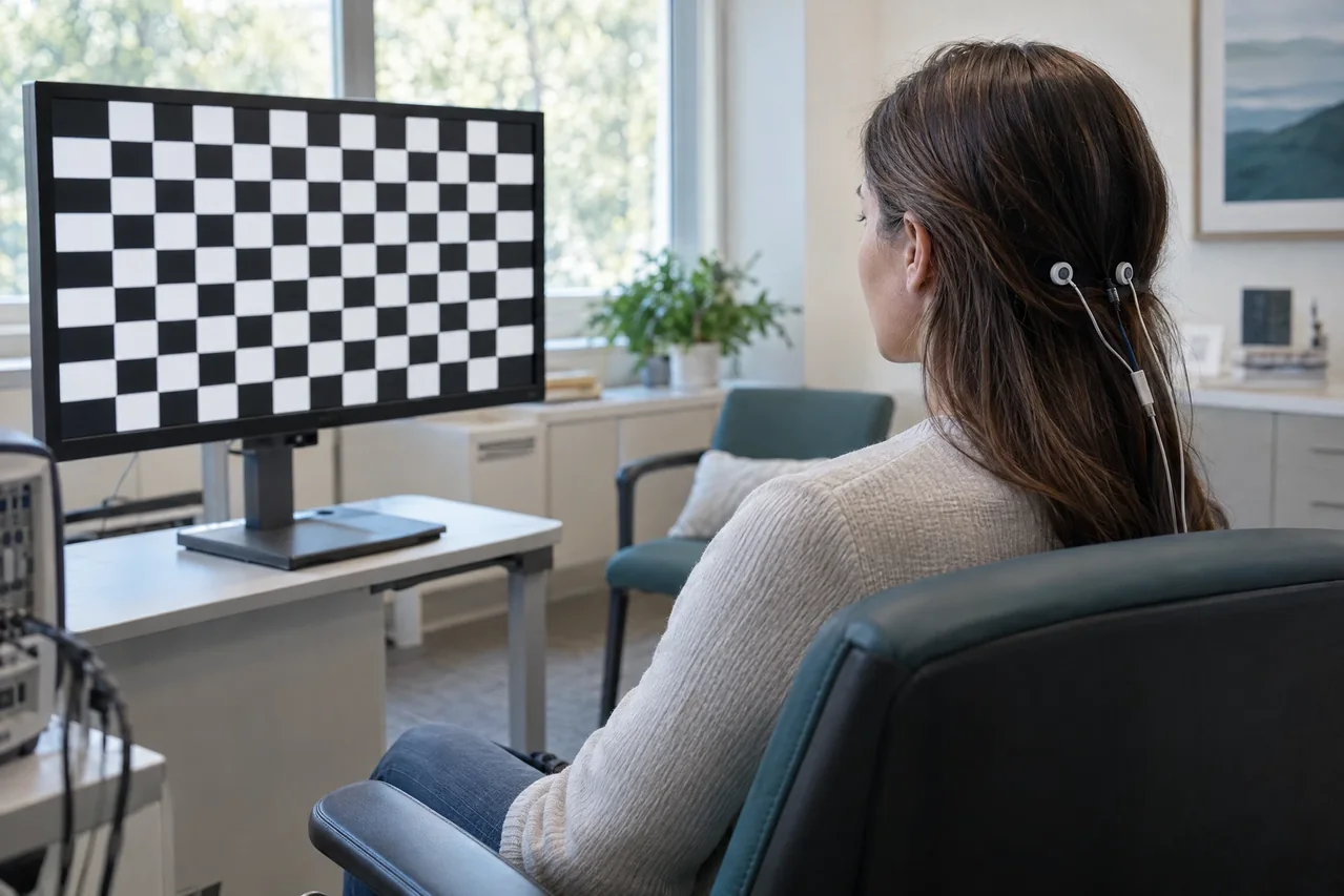
A visual stimulus, commonly a reversing checkerboard or, when fixation is limited, a flash, is presented while scalp electrodes record voltage over occipital regions. Repeated responses are averaged to reduce unrelated electroencephalographic noise and reveal reproducible stimulus-linked waveforms. Pattern, field, eye, and channel choices shape the physiologic question.
The response reflects synchronized activity as visual information travels from the retina through the optic nerves, chiasm, tracts, radiations, and visual cortex. Pattern-reversal recordings evaluate timing, polarity, and reproducibility of components such as N75, P100, and N145; P100 is a waveform landmark, not a universal diagnostic cutoff. Fixation, acuity, correction, attention, pupil and retinal factors, and technical quality can alter results.
Common clinical applications
Clinicians may request VEPs as an adjunct when evaluating suspected optic nerve dysfunction, demyelinating or inflammatory pathway disease, compressive or traumatic involvement, unexplained visual loss, or selected pediatric and neuro-ophthalmic questions. The test can provide functional evidence when routine eye assessments do not fully answer the question.
Pattern VEPs are generally more repeatable with adequate fixation, whereas flash VEPs can help when fixation or attention is difficult. VEPs may support assessment over time, but the indication and significance depend on clinical context and licensed medical judgment.
What you may expect
You will usually sit comfortably and look at a screen while small electrodes are attached to the scalp. One eye may be covered while the other views a checkerboard or light stimulus, then the eyes may be tested in turn. Skin cleaning, paste, or adhesive can feel mildly irritating; no test can guarantee no discomfort.
Keep your gaze on the target, remain as alert and still as possible, and report difficulty seeing or maintaining fixation. Setup and repeated trials take variable time; immediate results, a fixed duration, or a particular outcome should not be assumed.
For general preparation, bring usual glasses or contact lenses if used and arrive with clean, dry hair without oils, gels, or sprays so electrodes can make contact. Follow the ordering team’s instructions and tell the testing professional about relevant eye, skin, neurologic, or medical issues. Do not stop, change, or skip medications unless the clinician responsible for your care gives individualized instructions.
Explore the technical detail.
Open the sections below for anatomy, physiologic parameters, methodology and clinical limitations.
Anatomical & physiologic context
VEPs sample the visual system from retinal input and optic nerves through the optic chiasm and retrochiasmal pathways to occipital cortex. Monocular testing and, when indicated, multiple occipital channels can help compare eyes and describe response topography, while full-field stimulation is weighted toward central vision and may not expose every partial field lesion.
Important physiologic parameters
- Component latency: elapsed time from stimulus onset or reversal to identifiable waveform peaks, especially P100 when a pattern response is obtained.
- Amplitude and waveform morphology: voltage size, polarity, shape, and reproducibility, interpreted with signal quality and the stimulus protocol.
- Inter-eye and side-to-side relationships: relative timing, amplitude, and topographic differences under comparable conditions.
- Stimulus configuration: pattern versus flash, check or field size, contrast, luminance, reversal or presentation rate, monocular versus binocular delivery, and viewing distance.
- Recording conditions: montage, channels, filters, averaging, artifact rejection, fixation, alertness, visual acuity, correction, and ocular factors.
General methodology
A qualified testing professional checks vision-related conditions, prepares small scalp areas, applies surface electrodes, presents the selected stimulus to one or both eyes according to protocol, and records repeated trials while monitoring fixation, alertness, artifacts, and reproducibility. The study may be adapted for age, cooperation, visual acuity, and the referring clinical question.
Acquisition produces physiologic data; licensed medical interpretation, diagnosis and treatment decisions are separate clinical responsibilities. The recorded cortical response must be considered with visual, ophthalmic and neurologic information rather than interpreted as a standalone disease marker.
Clinical relevance & limitations
Interpretation weighs latency, amplitude, morphology, reproducibility, inter-eye differences, and topography against protocol-specific reference data and effects of acuity, refraction, fixation, attention, age, ocular disease, and artifact. A delayed, small, asymmetric, or absent response has multiple explanations and cannot identify cause without clinical correlation.
VEP data complement history, visual and neurologic examination, imaging, ophthalmic assessment, and other studies; they never establish a definitive diagnosis by themselves. Apex NeuroMetrics’ data-acquisition role, where applicable, does not confer authority to diagnose or treat. A licensed medical professional must interpret findings and determine their clinical significance within the full record.
- Normative limits and waveform labels vary with stimulus, montage, equipment, laboratory methods, patient factors, and visual conditions; no isolated latency, amplitude, or cutoff should be treated as a stand-alone diagnosis.
- A technically limited or normal VEP does not exclude visual pathway disease or independently determine prognosis, treatment, or symptom source.
Information, within appropriate scope.
Recorded physiologic data support broader clinical evaluation. Licensed medical interpretation, diagnosis and treatment are separate clinical responsibilities. Specific service availability, professional qualifications and arrangements must be verified.
Educational references
Selected sources support the educational discussion. They do not imply affiliation, endorsement or an individual clinical recommendation.