
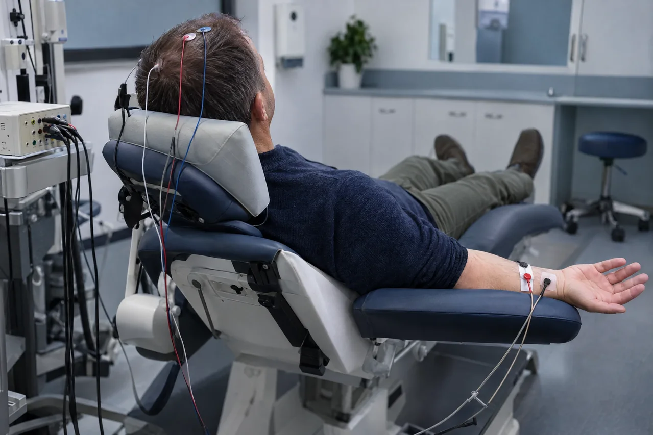
The study & its electrophysiologic principle
A brief stimulus is delivered repetitively at the wrist or ankle while surface electrodes record responses at peripheral, spinal, and scalp sites. Averaging trials improves signal-to-noise ratio and separates reproducible evoked activity from background.
SSEP waveforms reflect synchronized large-fiber somatosensory activity. Interpretation considers timing, size, configuration, side-to-side comparison, and reproducibility against reference data. Neural, technical, anesthetic, physiologic, and environmental factors can change results.
Common clinical applications
SSEPs may complement evaluation of sensory complaints, suspected spinal-cord or central sensory-pathway dysfunction, and disorders where objective pathway information is useful. They may contribute to functional localization or prognosis in defined contexts.
In specialized clinical settings, SSEP methods may also be used for selected intraoperative monitoring questions. That application involves a separate clinical, anesthetic and technical framework and is described for educational context—not as a claim that intraoperative monitoring is offered by Apex NeuroMetrics.
What you may expect
Skin electrodes and brief pulses may feel like tapping, tingling, or a quick twitch. Scalp preparation may involve gentle rubbing. The test is generally well tolerated, but discomfort and skin irritation are possible; no procedure guarantees a pain-free experience.
You may lie still with eyes closed while each limb is tested several times. Tell the team about implanted electrical devices or skin sensitivity. Do not stop prescribed medicines unless the clinician managing them instructs you to do so.
General preparation often includes clean skin and hair without lotions, oils, or styling products and comfortable clothing. Eating, drinking, duration, result timing, and arrangements vary; follow referring-service instructions.
Explore the technical detail.
Open the sections below for anatomy, physiologic parameters, methodology and clinical limitations.
Anatomical & physiologic context
Signals travel from the limb through a peripheral nerve and plexus, enter the spinal cord, ascend chiefly in the dorsal column–medial lemniscus system, relay subcortically, and reach contralateral primary somatosensory cortex. Montages may sample peripheral, cervical or lumbar, subcortical, and cortical components.
SSEP primarily informs large-fiber somatosensory pathway function. Preservation of a sensory response does not establish normal motor-pathway function or exclude all clinically relevant disease.
Important physiologic parameters
- Latency: stimulus-to-peak timing, interpreted with limb length, temperature, age, and laboratory references.
- Amplitude: waveform voltage; more variable than latency and judged with technical quality and reproducibility.
- Morphology and phase: component shape, polarity, and identifiable peaks help establish a genuine, comparable response.
- Side-to-side and segmental comparison: asymmetry or altered progression may help localize dysfunction, without constituting a cutoff diagnosis.
- Reproducibility and signal-to-noise ratio: repeated averages, stable contact, and controls support confidence.
- Context: stimulation, impedance, temperature, anesthesia, blood pressure, movement, artifact, and montage influence results.
General methodology
For a nonoperative study, a qualified testing professional places electrodes at appropriate scalp and selected peripheral or spinal recording sites, stimulates one limb at a time and acquires repeated averaged responses while the patient relaxes. Reproducibility, stimulus adequacy and artifact control are checked.
Technical acquisition produces waveforms and measurements; licensed medical interpretation is separate. SSEP findings must be considered by appropriately qualified healthcare professionals within the relevant clinical and professional scope.
Clinical relevance & limitations
SSEP findings are most useful alongside symptoms, history, neurologic examination, imaging, laboratory information, and other studies. An abnormality may indicate pathway dysfunction but does not alone identify cause or determine treatment.
The acquisition service records data; it does not independently diagnose, prescribe, or decide treatment. A licensed medical professional must interpret the study in context.
- Sensitivity and waveform quality vary with lesion location, peripheral neuropathy, temperature, edema, anesthesia, medications, artifact, and anatomy; a limited or normal study cannot exclude every important disorder.
- SSEP is not standalone and is not interchangeable with NCS, needle EMG, MEPs, F-waves, H-reflexes, or blink-reflex testing. No universal numeric cutoff or isolated change is definitive without qualified interpretation.
Information, within appropriate scope.
Recorded physiologic data support broader clinical evaluation. Licensed medical interpretation, diagnosis and treatment are separate clinical responsibilities. Specific service availability, professional qualifications and arrangements must be verified.
Educational references
Selected sources support the educational discussion. They do not imply affiliation, endorsement or an individual clinical recommendation.
- Recommendations of the International Society of Intraoperative Neurophysiology for intraoperative somatosensory evoked potentials (opens in a new tab)
- Guideline 11B: Recommended Standards for Intraoperative Monitoring of Somatosensory Evoked Potentials (opens in a new tab)
- Somatosensory evoked potentials (SSEP): Cambridge University Hospitals patient information (opens in a new tab)
- Recommendations of the International Society of Intraoperative Neurophysiology for intraoperative somatosensory evoked potentials: European Academy of Neurology summary (opens in a new tab)