
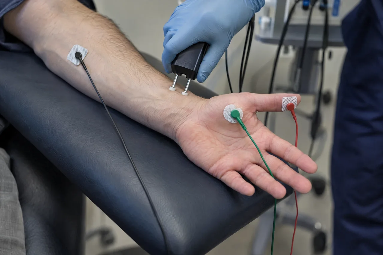
The study & its electrophysiologic principle
Surface stimulating electrodes deliver brief, controlled electrical pulses to a nerve, while recording electrodes detect a sensory response or the compound response of an innervated muscle. Stimulating at more than one site and relating response timing to measured distance helps characterize conduction along a nerve segment.
NCS primarily samples large myelinated sensory and motor fibers. Myelin supports rapid saltatory conduction, whereas axonal loss generally reduces responding fibers. Temperature, distance measurement, electrode placement, and technical factors influence recordings, so results require appropriate technique and clinical correlation.
Common clinical applications
Clinicians may use NCS in evaluating numbness, tingling, weakness, or persistent limb pain. Common questions include focal entrapment or compression, generalized peripheral neuropathy, and selected disorders affecting nerve roots or proximal segments.
NCS can help characterize sensory or motor involvement, axonal or myelin-related physiology, and focal or diffuse patterns. These are physiologic impressions, not stand-alone disease labels, and interpretation follows the clinical question.
What you may expect
You will usually sit or lie down while adhesive electrodes are placed on the skin. A brief pulse may feel like tingling, a tap, or a startle; sensations vary, and discomfort cannot be guaranteed to be absent. Tell the examiner if it becomes difficult to tolerate.
Preparation usually includes clean, lotion-free skin and loose clothing permitting access to the test areas. Do not stop or change medications unless the clinician managing them instructs you to do so. Tell the testing team about an implanted electrical device or blood-thinning medicines.
The number of nerves and sites determines duration, and NCS may be performed alone or before other electrodiagnostic components. A licensed medical professional must review findings and communicate their clinical meaning through the appropriate care pathway; immediate results and outcomes are not promised.
Explore the technical detail.
Open the sections below for anatomy, physiologic parameters, methodology and clinical limitations.
Anatomical & physiologic context
Studies may examine sensory, motor, or mixed peripheral nerves in the limbs and, when indicated, selected cranial pathways. A motor study records a muscle response after its supplying nerve is stimulated; a sensory study records the propagated response directly from the nerve. The segment tested is defined by stimulation and recording sites.
Optional late-response studies extend the physiologic view. An F-wave is a late motor response elicited by supramaximal stimulation, involving antidromic motor axon activation and subsequent motor-neuron backfiring; it is not a reflex. An H-reflex predominantly reflects a monosynaptic afferent–spinal–efferent arc. Blink-reflex testing samples trigeminal sensory input, facial motor output, and their brainstem circuitry.
Important physiologic parameters
- Latency: time from stimulation to the recorded onset or peak, interpreted according to the protocol.
- Response amplitude: the size of a sensory nerve action potential (SNAP) or compound muscle action potential (CMAP), influenced by responding fibers and technical factors.
- Conduction velocity: calculated from distance and latency differences between stimulation sites; it is not simply the latency at one site.
- Waveform and duration: morphology and temporal dispersion provide information about synchrony along the tested segment.
- Late responses: F-wave occurrence and latency, H-reflex recruitment and latency, or blink-reflex response timing may add information about proximal or cranial pathways when clinically appropriate.
General methodology
The examiner positions the patient, prepares clean skin, places electrodes, measures relevant distances, and delivers brief pulses while the instrument captures responses. Multiple nerves and sites may be tested; limb temperature and technical quality affect conduction measurements.
NCS is distinct from needle electromyography. Needle EMG examines electrical activity within muscle and requires performance by an appropriately qualified licensed professional; NCS data acquisition does not imply independent authority to perform needle EMG, interpret a complete electrodiagnostic examination, diagnose, or prescribe treatment.
Clinical relevance & limitations
Amplitudes, latencies, velocities, and waveforms complement the history, neurologic examination, and sometimes imaging, laboratory, genetic, or other electrodiagnostic studies. Values require suitable reference methods and clinical context.
Apex NeuroMetrics describes data acquisition and educational information. Licensed medical interpretation, diagnosis, and treatment decisions belong to the responsible qualified healthcare professionals and are outside what raw NCS data can establish by itself.
- NCS cannot establish a definitive diagnosis or replace history, examination, and other testing; reference expectations vary with age, body region, temperature, anatomy, and protocol.
- This educational description is not individualized medical advice and does not represent a guarantee of availability, interpretation arrangements, diagnosis, treatment, timing, or outcome.
Information, within appropriate scope.
Recorded physiologic data support broader clinical evaluation. Licensed medical interpretation, diagnosis and treatment are separate clinical responsibilities. Specific service availability, professional qualifications and arrangements must be verified.
Educational references
Selected sources support the educational discussion. They do not imply affiliation, endorsement or an individual clinical recommendation.
- American Association of Neuromuscular & Electrodiagnostic Medicine: Patient Information (opens in a new tab)
- Cleveland Clinic: Nerve Conduction Study (opens in a new tab)
- MedlinePlus: Electromyography (EMG) and Nerve Conduction Studies (opens in a new tab)
- Johns Hopkins Medicine: Nerve Conduction Studies (opens in a new tab)
- Jerath N, et al. F wave, A wave, H reflex, and blink reflex (opens in a new tab)