
The study & its electrophysiologic principle
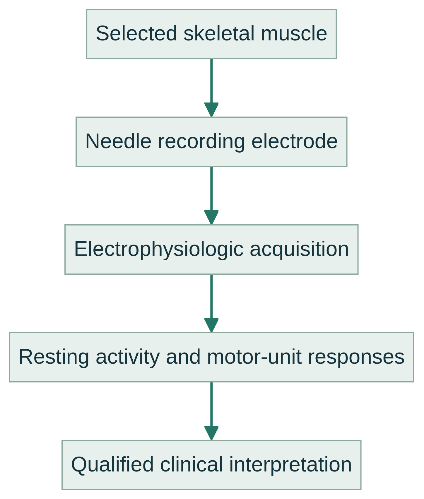
A sterile recording needle electrode is placed in selected muscles. The examiner observes insertional activity, activity while relaxed and motor-unit activity during graded contraction. Signals appear as waveforms and may be monitored acoustically; the needle records local muscle activity and does not deliver the nerve stimulus used for a nerve conduction study.
A motor unit comprises a motor neuron, its axon, neuromuscular junctions and supplied muscle fibers. Needle recordings sample nearby fibers. Spontaneous activity, motor-unit action-potential morphology and recruitment can reflect altered excitability, motor-unit loss or remodeling, but meaning depends on distribution, timing, technique and clinical context.
Common clinical applications
Qualified clinicians may use needle EMG to help evaluate suspected disorders affecting motor roots, plexuses, peripheral nerves, motor neurons, neuromuscular transmission or skeletal muscle, including weakness, atrophy, cramps or involuntary activity.
It can help localize and characterize a physiologic pattern and complement nerve conduction studies, imaging, laboratory testing and other assessments, without implying that every condition is detectable or a result alone confirms one disease.
What you may expect
Most people remain awake, seated or lying down, while several muscles are examined. Each placement may feel like a brief pinch or pressure, and contraction can cause discomfort; pain varies, so a pain-free test is not guaranteed. Equipment may make popping or crackling sounds.
Wear clothing permitting access and keep test areas free of lotions, creams or oils. Tell the examiner about anticoagulants or bleeding conditions, implanted electrical devices, medical conditions and all medicines or supplements. Do not stop medicines unless the prescriber gives individualized instructions.
A tested muscle may be tender or bruised briefly. Follow the clinical team’s instructions and report symptoms. Duration, result timing and next steps vary; immediate results and a particular outcome should not be assumed.
Explore the technical detail.
Open the sections below for anatomy, physiologic parameters, methodology and clinical limitations.
Anatomical & physiologic context
Muscles are selected to sample relevant spinal roots, peripheral nerves, plexus territories and, when indicated, cranial or paraspinal regions. Accurate localization requires anatomical knowledge, positioning, palpation and selective activation.
Needle EMG assesses muscle and motor-unit input locally. It does not image a nerve, spinal cord, brain or muscle structure, and cannot alone establish the cause of an abnormality.
Important physiologic parameters
- Insertional and resting activity: after adequate relaxation, the examiner assesses the response to needle movement and spontaneous discharges such as fibrillation potentials, positive sharp waves, fasciculation potentials or complex repetitive discharges.
- Motor-unit action potentials: during graded activation, the examiner evaluates waveform amplitude, duration, configuration and stability; these features vary with muscle, age, electrode, sampling site and effort.
- Recruitment and activation: the examiner evaluates the number and firing rates of motor units and interference-pattern density as contraction increases, considering pain, weakness and cooperation.
- Distribution and sampling: the pattern of findings across selected muscles, root levels and peripheral nerve territories, interpreted in relation to timing and clinical context.
General methodology
An appropriately qualified, licensed professional determines the clinical question, scope, muscles and real-time adjustments. A sterile needle electrode is inserted through cleansed skin; the patient relaxes and activates selected muscles with graded effort. Needle EMG requires appropriately qualified licensed professional performance.
Needle EMG is distinct from nerve conduction studies, which use surface electrodes and electrical stimulation to measure nerve responses. Data acquisition is not medical interpretation: a licensed interpreter integrates findings with history and examination and provides any conclusion within applicable scope.
Clinical relevance & limitations
Needle EMG findings are evidence, not a verdict. The licensed interpreter considers symptom chronology, neurologic examination, prior illness or surgery, medications, technical limitations and complementary studies; abnormalities can have more than one explanation.
Test data complement the history, examination and other studies. Medical interpretation, diagnosis and treatment decisions require the responsible qualified healthcare professional; they cannot be established by a recording alone.
- Needle EMG is invasive and sampling-based; selection, effort, pain, edema, body habitus and timing after injury can alter interpretation. No cutoff value or isolated waveform should be overread.
- Needle EMG performance and medical interpretation belong to appropriately qualified licensed professionals. Nerve conduction studies and F-waves, H-reflexes and blink responses are distinct procedures or measurements.
Information, within appropriate scope.
Recorded physiologic data support broader clinical evaluation. Licensed medical interpretation, diagnosis and treatment are separate clinical responsibilities. Specific service availability, professional qualifications and arrangements must be verified.
Educational references
Selected sources support the educational discussion. They do not imply affiliation, endorsement or an individual clinical recommendation.
- American Association of Neuromuscular & Electrodiagnostic Medicine: Reporting the Results of Nerve Conduction Studies and Needle EMG (opens in a new tab)
- Johns Hopkins Medicine: Electromyography (EMG) (opens in a new tab)
- Cleveland Clinic: EMG (Electromyography) (opens in a new tab)
- National Library of Medicine MedlinePlus: Electromyography (EMG) and Nerve Conduction Studies (opens in a new tab)
- Kim D-S. Needle Electromyography. Annals of Clinical Neurophysiology (opens in a new tab)
- Menkes DL. Needle EMG muscle identification: A systematic approach to evaluating the needle examination (opens in a new tab)